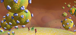
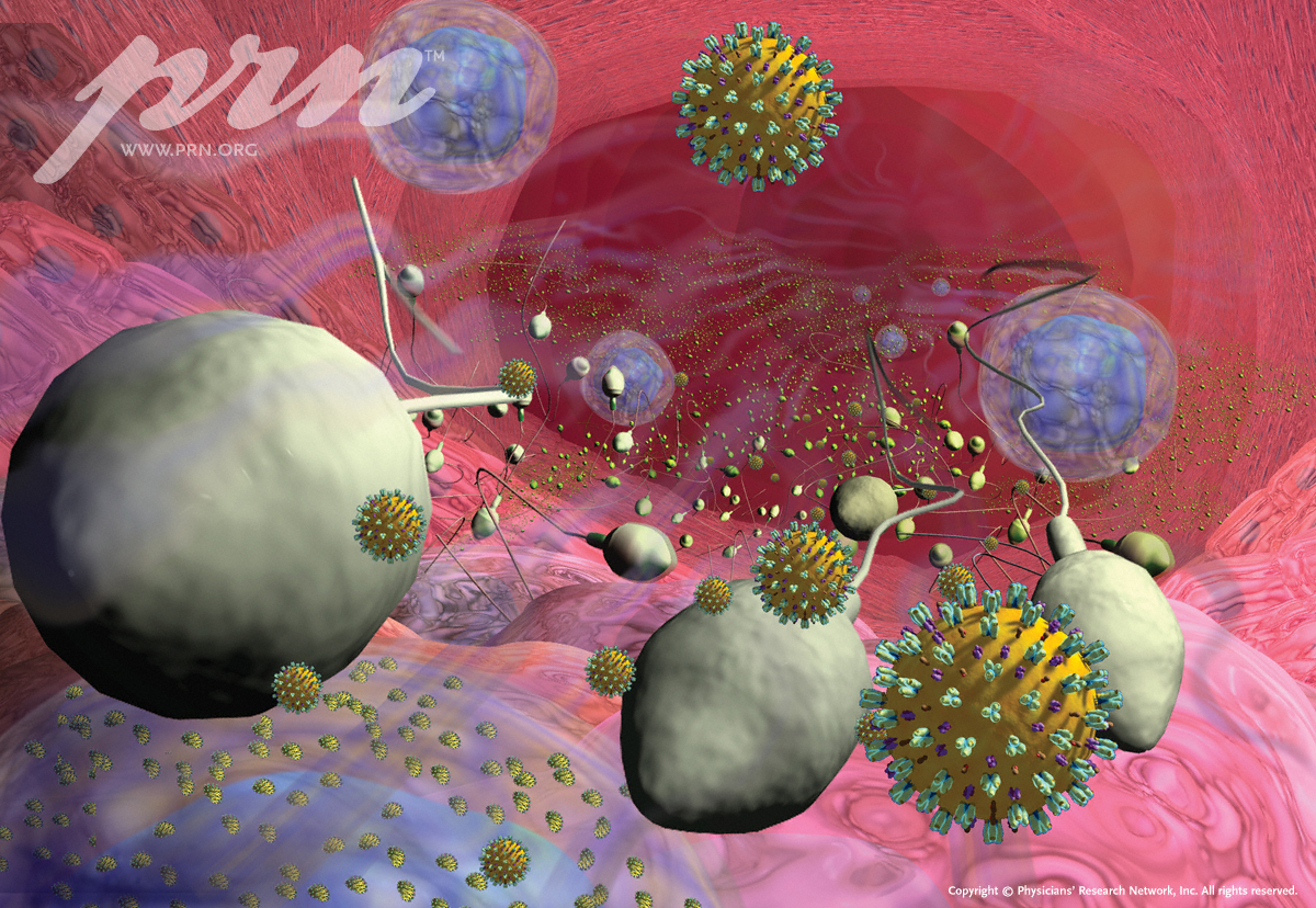
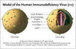
HIV Budding from Infected T-Lymphocytes (1200x800)
Our cover shows HIV budding from infected cells, in a new simulated model by Dr. Lou Henderson, that helps us visualize it better. The burden of HIV replication is never more profound than during the primary stage of HIV infection following transmission. And there is a resurgence of HIV from infected cells if treatment is not continuously suppressive.
Cover illustration by John Henderson based on virus models by L. E. Henderson, Ph.D, Frederick Cancer Research Center.
From: The PRN Notebook, Volume 6, Number 2; June 2001;
http://www.prn.org/
The PRN Notebook® is owned and published by Physicians¹ Research Network,® Inc. Copyright © 2001-2011, Physicians¹ Research Network, Inc. All rights reserved.
Download Wallpaper Image
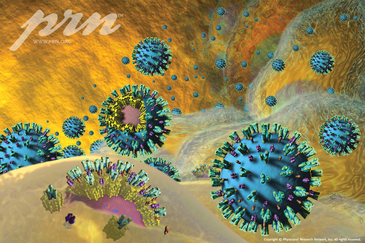
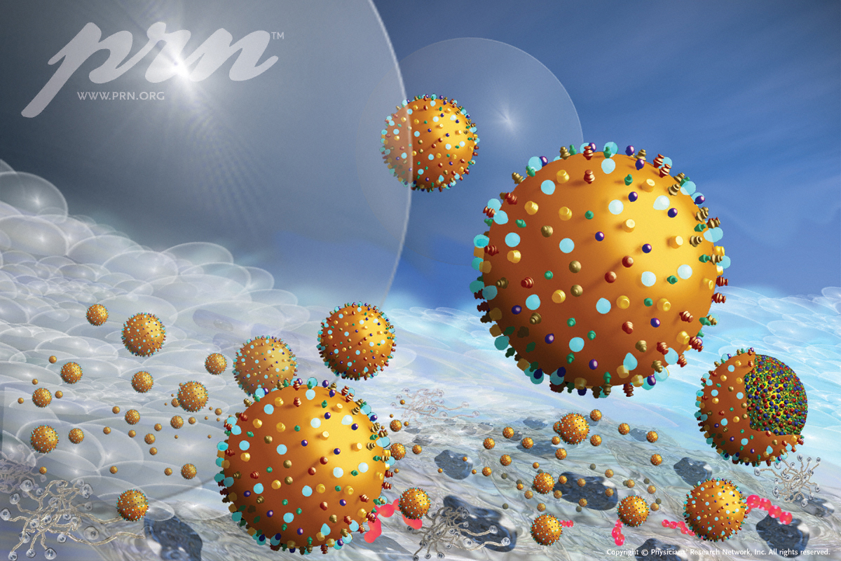
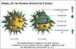
Model of the Human Hepatitis C Virus (1200x800)
Model of the Human Hepatitis C Virus
Illustration by John Henderson based on HCV models by Dr Louis Henderson, Frederick Cancer Research Center.
From: The PRN Notebook, Volume 6, Number 1; March 2001;
http://www.prn.org/
The PRN Notebook® is owned and published by Physicians¹ Research Network,® Inc. Copyright © 2001-2011, Physicians¹ Research Network, Inc. All rights reserved.
Download Wallpaper Image
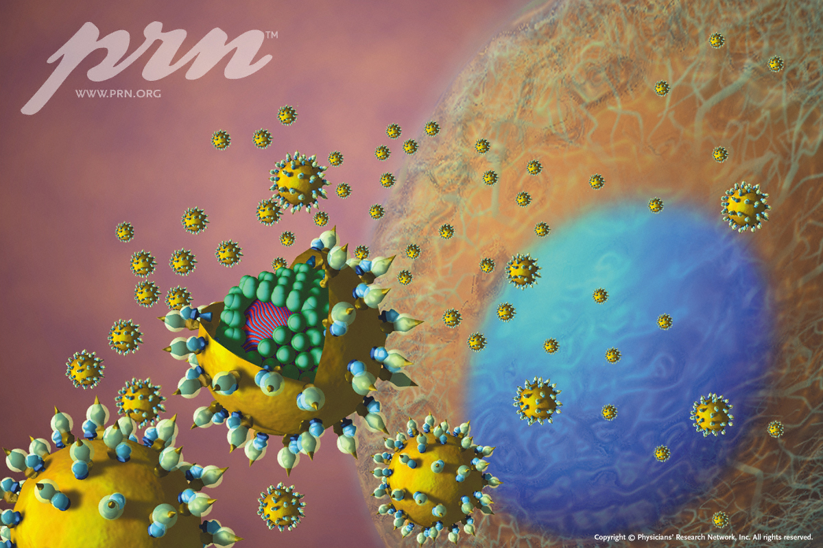
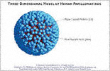
Oncogenic Human Papillomavirus (HPV) Shedding from Cervical or Anal Epithelial cells (1200x802)
Oncogenic human papillomavirus (HPV) shedding from cervical or anal epithelial cells.
The cover illustration is designed by Dr. Louis E. Henderson. Ph.D., based on a three-dimensional mpdel of the human papillomavirus.
From: The PRN Notebook, Volume 6, Number 3; September 2001;
http://www.prn.org/
The PRN Notebook® is owned and published by Physicians¹ Research Network,® Inc. Copyright © 2001-2011, Physicians¹ Research Network, Inc. All rights reserved.
Download Wallpaper Image
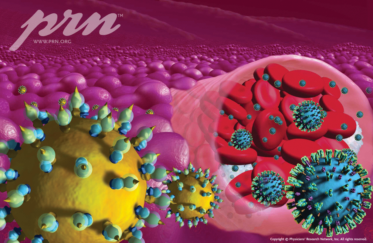
HIV in Semen: Appreciating the Risk (1200x828)
Semen is a complex mixture of fluids and cells from several organs including the testis, epididymis, seminal vesicles, prostate, and urethra. On average, ejaculated semen specimens contain100 million sperm. Also found in a single specimen of ejaculate are between 1 million and 10 million nonspermatozoal cells, including immature germ cells and leukocytes (e.g., CD4+ cells, CD8+ cells and monocytes/macrophages).
HIV has been detected in seminal leukocytes, but is probably not in spermatozoa themselves. The source of nonspermatozoal cells harboring HIV is still a matter of debate. One hypothesis is that HIV-infected leukocytes in semen arise from peripheral circulation via the prostate and seminal vesicles. Another hypothesis is that free virus infects germ compartment host cells, probably early in infection, leading to an immune-privileged, isolated reservoir of HIV infection and reproduction.
Viral load levels in the blood and genital fluids are often very high immediately following exposure, during a stage known as primary HIV infection (PHI). As is discussed in this issue of The PRN Notebook, there is evidence to suggest that newly infected persons are more likely to transmit HIV than those at subsequent stages of the disease, especially during the ³window period² before seroconversion. It is thus believed that transmission from people in the earliest stages of HIV disease may play a major role in the progression of the epidemic.
The cover illustration, is designed by Louis E Henderson, Ph.D.
From: The PRN Notebook, Volume 6, Number 4; December 2001;
http://www.prn.org/
The PRN Notebook® is owned and published by Physicians¹ Research Network,® Inc. Copyright © 2001-2011, Physicians¹ Research Network, Inc. All rights reserved.
Download Wallpaper Image
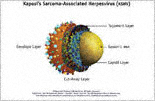
Kaposi’s Sarcoma-Associated Herpesvirus (KSHV), or Human Herpesvirus-8 (HHV-8), in Saliva (1200x800)
Saliva is a major source of infectious virus for many herpesviruses, including EBV and CMV, as well as KSHV; and by analogy to other saliva borne herpesviruses, KSHV in saliva is a likely source for transmission.
The cover illustration, as well as the three-dimensional model of KSHV, were created by Louis E Henderson, Ph.D.
From: The PRN Notebook, Volume 7, Number 1; March 2002;
http://www.prn.org/
The PRN Notebook® is owned and published by Physicians¹ Research Network,® Inc. Copyright © 2002-2011, Physicians¹ Research Network, Inc. All rights reserved.
Download Wallpaper Image
Liver tissue coinfected with HCV and HIV (1229x800)
Liver tissue coinfected with HCV and HIV.
Image and virus models created by Louis E Henderson Ph.D.
From: The PRN Notebook, Volume 11, Number 4; May 2007;
http://www.prn.org/
The PRN Notebook® is owned and published by Physicians¹ Research Network,® Inc. Copyright © 2007-2011, Physicians¹ Research Network, Inc. All rights reserved.
Download Wallpaper Image
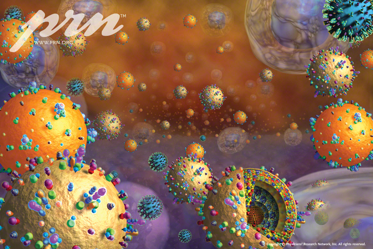
EBV and KSHV, in HIV Disease (1200x800)
In the setting of HIV disease, Epstein-Barr Virus (EBV) and Kaposi’s Sarcoma-associated Herpesvirus (KSHV/HHV-8) may transform B cells into lymphomas. KSHV is widely believed to be the causative agent of Kaposi’s sarcoma (KS) in individuals with immune suppression. It is also associated with the development of multicentric Castleman’s disease (MCD) and primary effusion (body cavity-based) lymphomas (PEL). EBV, an almost identical gammaherpesvirus, has been linked to a higher incidence of immunoblastic lymphoma and a heightened risk of primary central nervous system lymphoma. There is also some concern that loss of immune control of both herpesviruses might further increase the risk of these malignancies in AIDS.
The cover illustration, as well as the three-dimensional models of HIV, EBV and KSHV were created by Louis E. Henderson, PhD, Frederick Cancer Center.
From: The PRN Notebook, Volume 7, Number 3; September 2002; http://www.prn.org/
Download Wallpaper Image
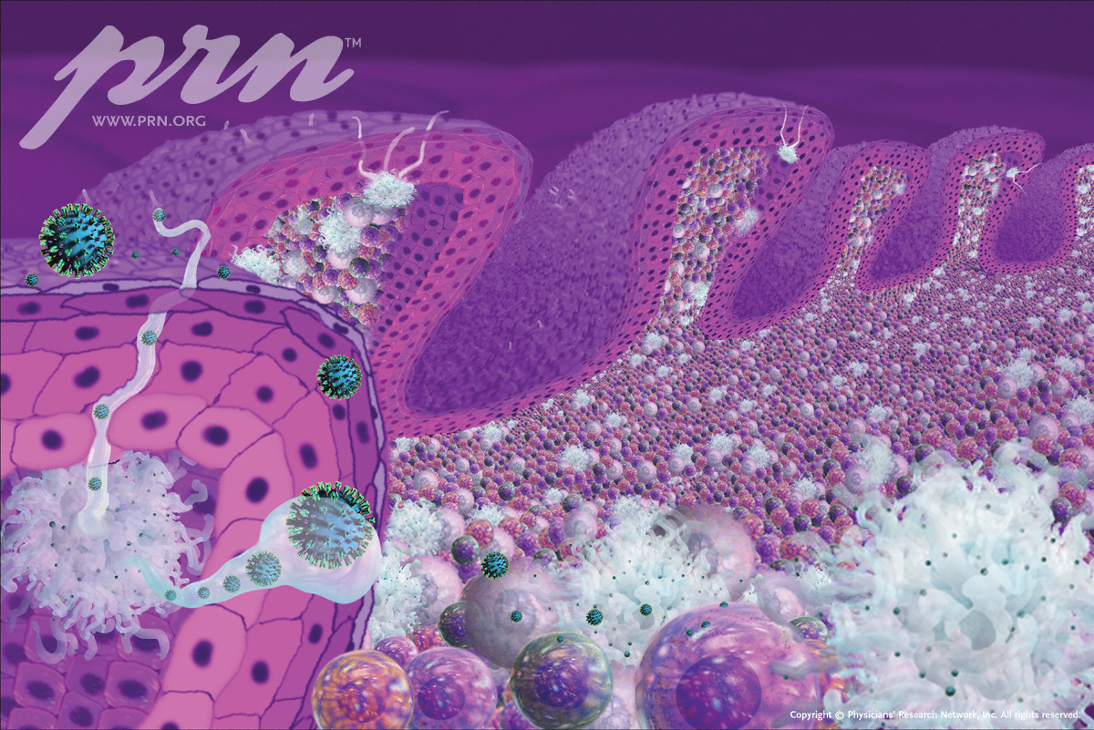
HIV in Mucosa-Associated Lymphoid Tissues (MALT) (1200x801)
Mucosal surfaces, including those in the genitourinary and gastrointestinal tracts, are important routes by which HIV may gain access to blood and lymphoid tissue during sexual transmission.
The cover illustration reflects one of several hypotheses surrounding HIV infection and replication in mucosa‹that dendritic cells can weave through mucosal tissue to engulf HIV at the surface and facilitate the presentation of virus or antigen to other lymphoid cells, such as macrophages, T-cells, and B-cells, in deeper layers. Understanding the precise mechanisms of viral transmission that occur in MALT may lead to new treatment options, better topical microbicides, and novel vaccine approaches.
The cover illustration, along with the HIV and cell models shown on this page, were created by Louis E. Henderson, Ph.D, Frederick Cancer Center
From: The PRN Notebook, Volume 7, Number 4; December 2002;
http://www.prn.org/
The PRN Notebook® is owned and published by Physicians¹ Research Network,® Inc. Copyright © 2002-2011, Physicians¹ Research Network, Inc. All rights reserved.
Download Wallpaper Image
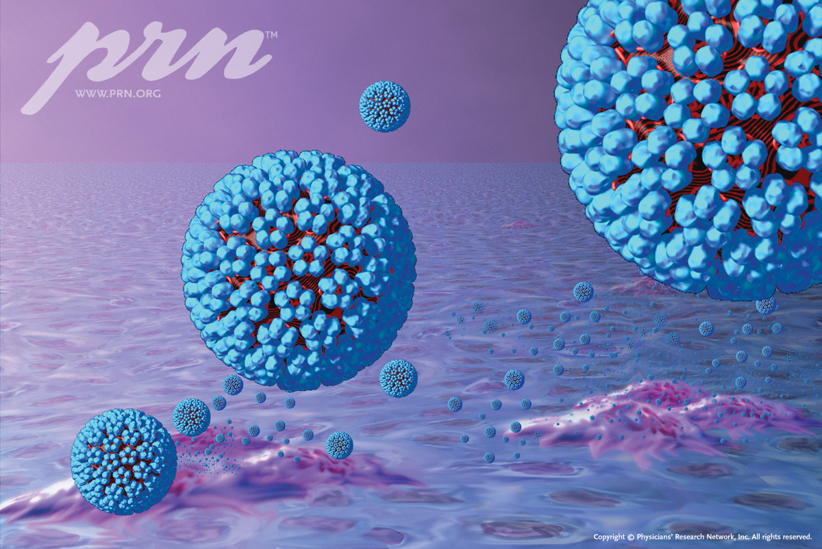
Human Immunodeficiency Virus (HIV) in Lymphoid Tissue (Model) (1200x1200)
Model of the Human Immunodeficiency Virus (HIV) in Lymphoid Tissue
Created by Louis E Henderson, Ph.D, of the Frederick Cancer Research Center.
From: The PRN Notebook, Volume 11, Number 3; November 2006;
http://www.prn.org/
The The PRN Notebook® is owned and published by Physicians¹ Research Network,® Inc. Copyright © 2006-2011, Physicians¹ Research Network, Inc. All rights reserved.
Download Wallpaper Image
M. tuberculosis bacilli in pools of bloody sputum in airway of HIV coinfected person (1229x800)
Clusters of infectious Mycobacterium tuberculosis bacilli in pools of bloody sputum in the airway of an HIV coinfected individual before coughing.
Mycobacterium tuberculosis (M. tb) is a bacteria transmitted via aerosolized droplet nuclei of sputum by individuals with pulmonary tuberculosis. TB is an AIDS defining coinfection and can be a life threatening complication of HIV disease, if not adequately treated.
Cover illustration created by Louis E. Henderson, Ph.D, of the Frederick Cancer Research Center.
From: The PRN Notebook, Volume 11, Number 2; October 2006;
http://www.prn.org/
The PRN Notebook® is owned and published by Physicians¹ Research Network,® Inc. Copyright © 2006-2011, Physicians¹ Research Network, Inc. All rights reserved.
Download Wallpaper Image
Community-Acquired Methicillin Resistant Staphylococcus Aureus (CA MRSA) and Pus in an Abscess
Community-acquired methicillin resistant Staphylococcus aureus (CA-MRSA) is a bacterium that causes painful skin infections and is associated with skin-to-skin contact. Poor HIV control, antibiotic usage, recreational substances and high-risk sexual behaviors may place HIV patients at heightened risk for CA-MRSA infections.
The cover illustration depicts CA-MRSA and pus in a skin and soft tissue infection of an HIV-infected individual, resulting in a painful and rapidly worsening abscess.
Created by Louis E Henderson, Ph.D, of the Frederick Cancer Research Center.
From: The PRN Notebook, Volume 11, Number 3; November 2006;
http://www.prn.org/
The PRN Notebook® is owned and published by Physicians¹ Research Network,® Inc. Copyright © 2006-2011, Physicians¹ Research Network, Inc. All rights reserved.
Download Wallpaper Image
Human Immunodeficiency Virus (HIV) 3-D Model with Cut Away
Hepatitis C Virus (HCV) 3-D Model With Cut-Away
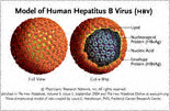
Hepatitis B Virus (HBV) 3-D Model With Cut-Away
Human Papillomavirus Virus (HPV) 3-D Model
Kaposi’s Sarcoma-Associated Herpes Virus (KSHV) 3-D Model
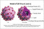
GB Virus-C (GBV-C) 3D Model with Cut-Away
|
|
Provider Resources
|